The rabbit monoclonal antibody R19M specifically binds to the scFv region of a CD19-specific mouse monoclonal antibody (mAb, clone FMC63). CD19 antigen is a B-cell specific cell surface antigen, which is expressed in all B-cell lineage malignancies and normal B-cells. The scFv region of FMC63 has been used to develop CD19-specific chimeric antigen receptor (CAR) T cells utilized in clinical trials.
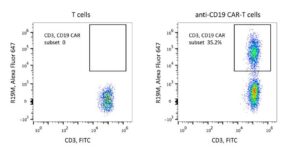
|
Item Information |
|
| Catalog # | 200102 |
| Size/Concentration | 100 tests |
| Price | $1,925.00 |
| Antibody Registration Number | AB_2857946 |
|
Specification |
|
| Host Species | Rabbit |
| Antibody Type | Monoclonal |
| Antigen Purification | Protein A |
| Fluorescent Dye | Alexa Fluor 647 |
| Excitation (nm) | 650 |
| Reactivity | Mouse |
| Immunogen | scFv region of a CD19-specific mouse mAb clone FMC63 |
| Storage Buffer | Aqueous buffered solution containing protein stabilizer and ≤0.03% sodium azide |
| Application | FCM |
Shipping and Storage
| Shipping | The product is shipped at 2-8°C. |
| Stability & Storage | To provide optimal stability of reagent:
|
FACS Protocol
- If you want to set a control, the recommended reagent to use is the following: Rabbit IgG Isotype Control, Alexa Fluor 647 (Product No. 700302).
(Optional) For Whole Blood Sample
1. Pipette 1 μL Rabbit Anti-Mouse C11D5.3 scFv Monoclonal Antibody, Alexa Fluor 647 into the bottom of the tube.
2. Add dead cell staining solution and additional fluorochrome conjugated antibodies into the bottom of the tube.
3. Pipette 100 µL of well-mixed, anticoagulated whole blood into the bottom of the tube. Mix gently and thoroughly.
Note Avoid smearing sample down the side of the tube. If the sample remains on the side of the tube, it will not be stained with the reagents.
4. Incubate for 25 minutes in the dark at room temperature (18-25°C).
5. Pipette Red Blood Cell Lysis Solution to the tube. Mix gently and thoroughly. Incubate for 15 minutes in the dark at room temperature (18-25°C).
6. Add 500 μL FACS buffer to the tube. Mix well and centrifuge at 300g for 5 minutes at room temperature (18-25°C). Aspirate supernatant
completely.
7. Repeat step 6 twice.
8. Add a suitable amount of FACS buffer to resuspend cell and analysis by flow cytometry.
(Optional) For Cell Sample
1. Harvest the cells and wash the cells twice by FACS buffer.
2. Count the cells number and the viability.
3. Resuspend the cell suspension to a concentration up to 1×10⁶ nucleated cells per 100 μL of buffer.
4. Add 1 μL Rabbit Anti-Mouse C11D5.3 scFv Monoclonal Antibody, Alexa Fluor 647, dead cell staining solution and additional fluorochrome. Mix
gently and thoroughly
5. Incubate for 25 minutes in the dark at room temperature (18-25°C).
6. Add 500 μL FACS buffer to the tube. Mix well and centrifuge at 300 g for 5 minutes at room temperature (18-25°C). Aspirate supernatant
completely.
7. Repeat step 6 twice.
8. Add a suitable amount of FACS buffer to resuspend cell and analysis by flow cytometry
Citations
- Agarwal S, Aznar MA, Rech AJ, Good CR, Kuramitsu S, Da T, Gohil M, Chen L, Hong SA, Ravikumar P, Rennels AK, Salas-Mckee J, Kong W, Ruella M, Davis MM, Plesa G, Fraietta JA, Porter DL, Young RM, June CH. Deletion of the inhibitory co-receptor CTLA-4 enhances and invigorates chimeric antigen receptor T cells. Immunity. 2023 Oct 10;56(10):2388-2407.e9. doi: 10.1016/j.immuni.2023.09.001. Epub 2023 Sep 29. PMID: 37776850; PMCID: PMC10591801.
- Cheng, Jiali, et al. Monitoring Anti‐CD19 Chimeric Antigen Receptor T Cell Population by Flow Cytometry and Its Consistency with Digital Droplet Polymerase Chain Reaction. Aug. 2022, https://doi.org/10.1002/cyto.a.24676.
- Cheng J, Yan J, Liu Y, Shi J, Wang H, Zhou H, Zhou Y, Zhang T, Zhao L, Meng X, Gong H, Zhang X, Zhu H, Jiang P. Cancer-cell-derived fumarate suppresses the anti-tumor capacity of CD8+ T cells in the tumor microenvironment. Cell Metab. 2023 Jun 6;35(6):961-978.e10. doi: 10.1016/j.cmet.2023.04.017. Epub 2023 May 12. PMID: 37178684.
- Cheng J, Mao X, Chen C, Long X, Chen L, Zhou J, Zhu L. Monitoring anti-CD19 chimeric antigen receptor T cell population by flow cytometry and its consistency with digital droplet polymerase chain reaction. Cytometry A. 2023 Jan;103(1):16-26. doi: 10.1002/cyto.a.24676. Epub 2022 Aug 5. PMID: 35875964; PMCID: PMC10087589.
- Lei W, Zhao A, Liu H, Yang C, Wei C, Guo S, Chen Z, Guo Q, Li L, Zhao M, Wu G, Ouyang G, Liu M, Zhang J, Gao J, Qian W. Safety and feasibility of anti-CD19 CAR T cells expressing inducible IL-7 and CCL19 in patients with relapsed or refractory large B-cell lymphoma. Cell Discov. 2024 Jan 9;10(1):5. doi: 10.1038/s41421-023-00625-0. PMID: 38191529; PMCID: PMC10774422.
- Wu W, Zhou Q, Masubuchi T, Shi X, Li H, Xu X, Huang M, Meng L, He X, Zhu H, Gao S, Zhang N, Jing R, Sun J, Wang H, Hui E, Wong CC, Xu C. Multiple Signaling Roles of CD3ε and Its Application in CAR-T Cell Therapy. Cell. 2020 Aug 20;182(4):855-871.e23. doi: 10.1016/j.cell.2020.07.018. Epub 2020 Jul 29. PMID: 32730808.

